Ontario Institute for Cancer Research / Thomas Hudson, MD.
McGill University / Robert Sladek, MD.
Center for Mathematical Biosciences / Bernhard Bodmann, Robert Azencott.
Data
:
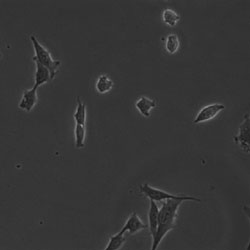
Bright field live-cell microscopy (20 views / hr)
Goal
:
Automatic extraction of cell components and fast phenotyping
Challenge
:
Diffraction patterns cause misclassifications
Mathematics
:
Develop a convolution-insensitive machine learning algorithm
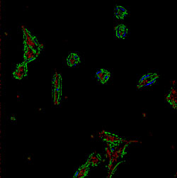
Left: Bright field image to be classified. Right: Output of a standard support-vector machine classifier.
Color coding corresponds to statistics of nucleus (red) cytoplasm (green), dividing cell (blue).